Tooth decay - symptoms and treatment
Definition of the disease. Causes of the disease
The causes of caries are reduced to two etiological factors:
Common factors:
Inadequate nutrition and diet. The lack of a whole complex of vitamins and minerals leads to a disorganization of the tooth structure. At the same time, the pathological effect of easily digestible carbohydrates, the nature and diet, and the fluorine content in drinking water play a huge role.
Somatic diseases that are directly related to the development and formation of the tooth and its elements, which can cause functional and structural changes.
The influence of damaging environmental factors, extreme impact on the macroorganism (overheating, frostbite, etc.).
Hereditary factors associated with the full formation of the structure and chemical composition of tooth tissues.
Local factors:
Dental plaque rich in microorganisms.
Exposure to the composition of the oral fluid, which may change as a result of various diseases.
The consistency of the local protective mechanisms of tooth tissues, their resistance (stability).
The condition of the pulp is the connective tissue filling the cavity of the teeth.
Normal laying and maturation of the dentition.
Deviations in the biochemical composition of tooth tissues.
Caries can occur at any age. Most often, it gradually appears in adults in 35-44 years. Children are especially prone to caries, since milk enamel is much more sensitive than molar enamel.
If you find similar symptoms, consult your doctor. Do not self-medicate - it is dangerous for your health!
Symptoms of tooth decay
It is important to understand that the appearance of caries can be both an acutely ongoing process and a chronically developing one. In the first case, we are talking about hereditary failure of the strength and stability of tooth tissues. A chronic process arises as a result of prolonged exposure to local and general damaging factors.
As a rule, the disease occurs gradually, as compensatory-adaptive mechanisms protect the body from unpleasant symptoms for a long time. However, at a certain point, they still fail, and the first signs of caries appear in the form of discomfort in the oral cavity when eating cold, hot, spicy, sour or sweet food. Further progression of the disease leads to the development of plaque and the appearance of tartar, worsening the condition of the oral cavity.
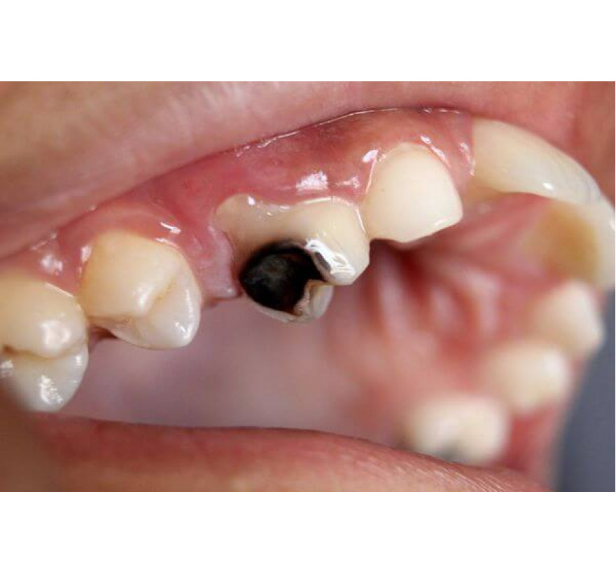
Another important symptom is the presence of carious spots, which at first are single, but gradually their number and area increase. The color of the tooth itself is not changed, with the exception of the affected area.
With a slow course of the carious process, the affected area is colored yellow or dark brown. With a rapid course of caries, which is more often observed in children due to increased permeability of tissues of milk teeth and the individual reactivity of the body, the affected areas are bright. The mucosa in the surrounding tooth tissue during caries is not changed, tapping on the tooth is painless.
The most unpleasant symptom of caries is bleeding gums. It also develops gradually: if at first the gums bleed during brushing, but, progressing, bleeding can occur spontaneously.
The clinic of carious lesions is also characterized by the occurrence of localized pain when exposed to an irritant, after the elimination of which the pain quickly subsides. This symptom occurs when tooth decay penetrates the dentin - the bone tissue of the tooth.
Pathogenesis of tooth decay
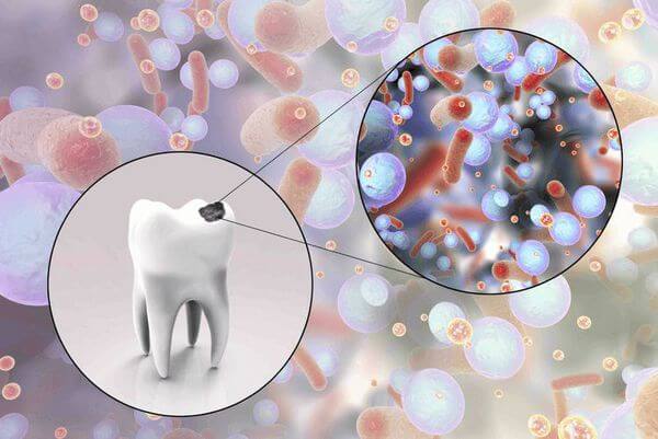
Currently, in medicine there are a huge number of theories of caries. The generally accepted is the multifactorial theory, according to which the demineralization of the hard tissues of the tooth proceeds under the influence of microorganisms that accumulate in the plaque. The result of their vital activity leads to the formation of organic acids that destroy the protective elements of tooth tissue.
The formation of caries is based on a carious situation, which gives rise to the formation of a carious spot.
As shown in the diagram, caries formation is possible with a single exposure to various factors. Only with their combined influence does the process of formation of dental plaque start, which gives rise to caries.
Stages of development of dental plaque:
Pellicle is an organic film that forms on the surface of a tooth within minutes and hours.
The growth of bacterial colonies - occurs during the first few days immediately after adsorption (absorption) on the surface of the pellicle of epithelial cells and microorganisms.
Stage mature plaque - gives rise to caries progression
The rate of development of this process directly depends on the nature of food and the quality of oral hygiene. That is why daily brushing is extremely important.
Classification and stages of tooth decay
The following varieties of caries are distinguished:
By the nature of the affected tooth:
decay of deciduous teeth;
caries of molar (permanent) teeth.
By age characteristics of occurrence:
juvenile caries - occurs at a young age;
senile caries - occurs in adulthood.
Depending on the initial link of the lesion:
primary caries - the formation of a carious defect on a healthy tooth enamel that has not previously been treated;
secondary caries - the occurrence of caries on a sealed treated tooth (carious lesion of depulled teeth and caries located under an artificial crown).
By the presence or absence of complications:
complicated caries - a carious process with the development of periodontitis and pulpitis;
uncomplicated caries.
By the number of affected teeth:
single caries - no more than five teeth are affected;
multiple caries - more than five teeth are involved in the carious process.
The concept of “blooming caries” is also distinguished - an acute process that affects most teeth.
Anatomists divide caries into:
tooth decay of the tooth;
tooth decay;
tooth decay.
Caries can be localized in different parts of the teeth, so there are four forms of caries:
fissure caries - carious lesion of the surface of the chewing teeth and premolars;
approximate caries - occurs between the teeth;
cervical caries - formed in the neck of the tooth;
circular caries - a carious lesion surrounding the neck of the tooth.
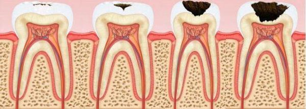
Depending on the depth of the lesion and the speed of the carious process, there are four forms of caries:
Initial caries or carious stain - no complaints; a chalky spot is found on the tooth surface.
Superficial caries - only tooth enamel is affected; complaints are either absent or pain from chemical and temperature irritants occurs.
Medium caries - enamel and the surface layer of dentin are affected; the pain reaction may not be expressed or may arise from eating and mechanical irritation when opening the carious cavity.
Deep caries - enamel and a significant layer of dentin are affected, but a thin layer remains above the cavity of the tooth; pain arises from the action of mechanical, chemical and temperature irritants; with a prolonged course of the carious process, pain symptoms are less pronounced.
Acute and chronic forms of caries are distinguished with the course. In this case, do not forget about the most acute and asymptomatic forms of the disease.
The World Health Organization also has its own classification, according to which caries is divided into:
initial caries;
enamel caries;
caries of dentin;
cement decay;
stopped caries (when the carious process is stabilized).
It is also worth noting that there are caries-susceptible zones. In this regard, caries may be:
typical - located in the natural recesses of the teeth, contact surfaces, as well as in the cervical areas;
atypical - it is most often localized in areas resistant to caries formation, for example: the equator, mounds of chewing teeth, oral surfaces of teeth, in the area of the tooth root.
Complications of tooth decay
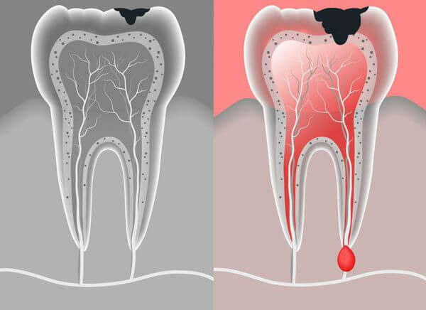
Quite often, patients ignore the first manifestations of caries, in connection with this, the disease progresses, carious plaque increases, and microorganisms penetrate deep into the tooth. As a result, there are complications that cannot always be cured immediately, since they require long-term treatment and the intervention of dentists of various specializations. Such complications include: pulpitis, periodontitis and granuloma.
One of the most common complications of late treatment to the doctor is pulpitis - diffuse inflammation of the tooth pulp, which is localized inside the cavity and gradually leads to thinning of dentin and enamel.
Further progression of pulpitis can lead to periodontitis - further inflammation that spreads to the bone tissue surrounding the tooth. In this case, the patient has general and local symptoms of inflammation:
fever;
swelling;
sharp throbbing pain when pressed;
gum swelling.
In this case, a visit to a doctor is necessary, since periodontitis can become the initial link in the process of acute suppurative inflammation, leading to osteomyelitis (bone inflammation) of the jaw and generalized infection of the whole organism - sepsis.
Chronically, current periodontitis can cause the formation of granulomas - small nodules filled with purulent masses. Their occurrence is an urgent indication for surgical treatment, since they can be transformed into a tooth cyst, and then phlegmon - spilled acute inflammation.
Unfortunately, not all patients understand the need for timely treatment of caries. However, complications of caries can sometimes lead to irreversible secondary changes and a deterioration in the quality of human life.
Diagnosis of tooth decay
As you know, timely diagnosis always contributes to a more favorable outcome of the disease. In the practice of the dentist there is a large arsenal of different methods for the differential diagnosis of caries. For this, general and additional methods are used.
General diagnostic methods:
Inspection and questioning of the patient. Despite its simplicity, it is of great importance and requires the care and knowledge of a doctor. The dentist examines with a mirror, carefully examining each tooth for cavities and stains, as well as the changed color of the tooth. During the interrogation, the dentist must find out what complaints the patient has addressed, find out about the presence of pain, under what conditions they arise and about their nature.
Percussion. Tapping allows the doctor to form an idea of the nature of the pain, its irradiation and localization, as well as to identify compaction, granulomas, etc.
Sounding. This diagnostic method is a more advanced inspection method. It is performed using a special device - a probe, which allows the doctor to more accurately establish the degree of caries and its localization.
Thermal test. It belongs to the most affordable diagnostic methods, as it is carried out using a jet of cold water or cotton balls soaked in special solutions. A thermal test allows you to understand the degree of tooth soreness and check the stability of dental tissue in response to a cold (most often) stimulus.
Additional diagnostic methods:
Electrodontometry. It represents the effect of an electric current on the pulp of a tooth, it is necessary to identify the state of the nerve endings of the tooth. The advantages of this method are minimal pain, but in practice it is rarely carried out.
Vital staining. This method was proposed by E.V. Borovsky. It is carried out as follows: the tooth is pre-cleaned and dried, then a dye is applied to it (most often methylene blue), as a result of which carious spots and cavities are stained.
Transluminescence. The study is carried out in a dark room, a dye is pre-applied to the teeth. Carious formations differ from healthy areas by the presence of dark spheres.
Luminescent method. It is based on the passage of ultraviolet light through the dental tissue. At the same time, healthy teeth remain bright, and those affected by caries become dark.
Determination of electrical resistance of hard tissues. Due to the loss of mineral components, tooth decayed teeth when exposed to electric current have less electro-resistance compared to healthy teeth.
X-ray diagnostics. It is one of the earliest and simplest studies. It allows you to identify structural changes in tooth tissues and hidden carious cavities.
Tooth decay treatment
Caries can be treated with invasive and non-invasive methods.
Non-invasive methods are certainly simpler for both the doctor and the patient. Their essence is remineralization (restoration of mineral balance) of tooth tissues. However, such treatment is possible only at the initial stages of caries development, when there are no visible defects, and only the dynamic balance between the content of organic acids and the mineral component of the tooth is broken.
Non-invasive methods include treatment with various preparations of calcium, fluorine and other mineral-containing substances. In practice, such drugs as calcium gluconate, remodent, calcium glycerophosphate, calcium hydroxide, sodium fluoride, fluorine-containing gel are most often used. The methods of their use are different: from rubbing a fluorine-containing gel to the introduction of calcium and fluorine preparations into tooth tissues using electrophoresis.
The course of remineralizing therapy consists of 15-20 procedures.
Also an important method of treatment is professional oral hygiene, which consists in cleaning necrotic masses and food residues of hard-to-reach areas of the oral cavity.
Invasive methods are resorted to in cases where the caries formation process spreads to deeper layers of tooth tissues. This treatment is called "preparation followed by filling."
This treatment pursues the following goals: removal of necrotic masses, application and fixation of fillings, restoration of the anatomical structure of the tooth. This process takes place in several stages:
opening of the carious cavity with the help of spherical and fissure boron;
expansion of the carious cavity to prevent secondary caries (most domestic authors adhere to the method of I.G. Lukomsky);
necrotomy with subsequent removal of the destroyed and softened dentin;
giving a cavity to the shape of a geometric figure;
preventive treatment of enamel edges;
antiseptic neutralization of the carious cavity;
dehydration with 96% alcohol;
filling.
Forecast. Prevention
The prognosis of caries depends on the depth of destruction that affected the tooth tissue, but in general it is favorable in connection with the development of treatment methods and timely diagnosis.
As you know, the best treatment is disease prevention. In particular, caries prophylaxis can be divided into endogenous (internal) and exogenous (external). [6]
Endogenous prophylaxis:
Proper nutrition, which includes all the necessary micro and macro elements. It is one of the fundamental principles of caries prevention, especially in children and in people with reduced body resistance.
The use of high-quality drinking water containing the necessary mineral salts.
The use of various drugs that strengthen the tooth tissue.
Eating the most important vitamins for dental tissue - A, B, C, D.
Exogenous prophylaxis:
Fluoroprophylaxis and remineralization.
The use of special preventive toothpastes.
Fissure sealing.
One of the most important aspects of the prevention of dental diseases is the rehabilitation (improvement) of the oral cavity. It includes several stages:
examination of the oral cavity;
treatment of both milk and permanent teeth;
elimination of sites affected by infectious agents;
treatment of affected mucous membranes;
correction and prevention of tooth and jaw deformation;
periodic and scheduled medical examinations.
Well, of course, it is worth remembering the most important thing: timely contact with a specialist will prevent from the adverse effects of caries.