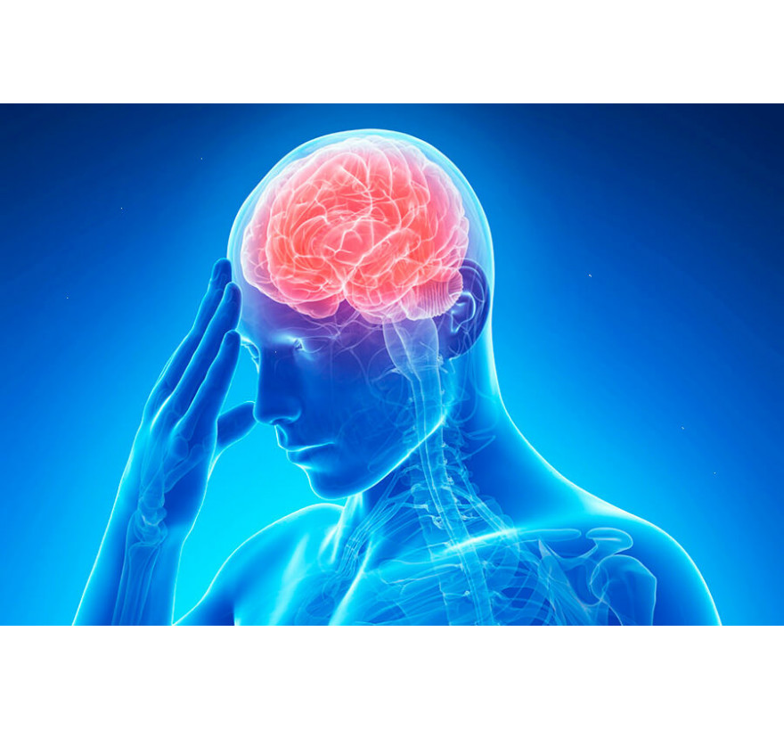
Stroke: symptoms, first signs, treatment
You can diagnose a stroke by the totality of data from clinical, laboratory, tomographic and vascular studies. Treatment consists of maintaining the body’s vital functions, correcting cardiac, respiratory and metabolic disorders, combating cerebral edema, specific pathogenetic, neuroprotective and symptomatic therapy, and preventing complications.
General information
Stroke is an acute vascular catastrophe resulting from vascular diseases or abnormalities of the vessels of the brain. In Russia, the incidence reaches 3 cases per 1,000 population. Strokes account for 23.5% of the total mortality of the Russian population and almost 40% of mortality from diseases of the circulatory system. Up to 80% of stroke patients have persistent neurological disorders causing disability. About a quarter of these cases are profound disabilities with a loss of self-care. In this regard, the timely provision of adequate emergency medical care for stroke and full rehabilitation are among the most important tasks of the healthcare system, clinical neurology and neurosurgery.
There are 2 main types of stroke: ischemic and hemorrhagic. They have fundamentally different development mechanisms and require radically different approaches to treatment. Ischemic and hemorrhagic strokes occupy 80% and 20% of the total set of strokes, respectively. Ischemic stroke (cerebral infarction) is caused by impaired cerebral artery patency, leading to prolonged ischemia and irreversible changes in brain tissue in the blood supply zone of the affected artery. Hemorrhagic stroke is caused by a pathological (atraumatic) rupture of the cerebral vessel with hemorrhage in the cerebral tissue. Ischemic stroke is more often observed in people older than 55-60 years of age, and hemorrhagic stroke is characteristic of a younger category of the population (usually 45-55 years).
Causes of stroke
The most significant factors in the occurrence of stroke are arterial hypertension, coronary heart disease and atherosclerosis. Unhealthy diet, dyslipidemia, nicotine addiction, alcoholism, acute stress, adynamia, and oral contraceptives contribute to the development of both types of stroke. In this case, malnutrition, dyslipidemia, arterial hypertension and adynamia do not have gender differences. Obesity is a risk factor that occurs mainly in women, and alcoholism in men. Increased risk of stroke in those individuals whose relatives suffered a vascular accident in the past.
Ischemic stroke develops due to a violation of the passage of blood through one of the blood vessels supplying the brain. And we are talking not only about intracranial, but also about extracranial vessels. For example, occlusion of the carotid arteries accounts for about 30% of cases of ischemic stroke. The cause of a sharp deterioration in cerebral blood supply can be vascular spasm or thromboembolism. The formation of thromboembolism occurs in cardiac pathology: after a myocardial infarction, with atrial fibrillation, acquired valve heart defects (for example, with rheumatism). Blood clots formed in the cavity of the heart move to the cerebral vessels, causing them to become clogged. The embolus may be a part of the atherosclerotic plaque that has torn off from the vascular wall and, when it enters the smaller cerebral vessel, leads to its complete occlusion.
The occurrence of hemorrhagic stroke is associated mainly with diffuse or isolated cerebral vascular pathology, due to which the vascular wall loses its elasticity and becomes thinner. Similar vascular diseases are: cerebral arteriosclerosis, systemic vasculitis and collagenoses (Wegener's granulomatosis, SLE, periarteritis nodosa, hemorrhagic vasculitis), vascular amyloidosis, angiitis with cocaine addiction and other types of drug addiction. Hemorrhage may be due to an abnormality with the presence of arteriovenous malformation of the brain. Changing the site of the vascular wall with loss of elasticity often leads to the formation of an aneurysm - protrusion of the artery wall. In the area of the aneurysm, the vessel wall is very thinned and easily ruptured. The gap contributes to a rise in blood pressure. In rare cases, a hemorrhagic stroke is associated with impaired blood coagulation with hematological diseases (hemophilia, thrombocytopenia) or inadequate therapy with anticoagulants and fibrinolytics.
The cause of this sudden condition is acute failure or complete cessation of blood supply to any area of the brain. This can occur due to a strong narrowing of the lumen of the vessel affected by atherosclerotic plaques, clogged arteries with a thrombus, or cholesterol deposition. In this case, I diagnose ischemic stroke (AI).
The diagnosis of hemorrhagic stroke (GI) is established when confirming rupture of the artery and hemorrhage in the substance of the brain. In neurological practice, in 75–85% of cases I diagnose ischemic strokes, hemorrhagic strokes account for 20–25%.
Etiological factors of ischemic brain damage are divided into two groups:
1. Unmodified:
age;
hereditary predisposition;
smoking;
excess body weight;
alcohol;
sleep apnea;
addiction;
frequent psycho-emotional overstrain;
lack of exercise.
2. Modified:
arterial hypertension;
damage to the carotid arteries;
dyslipoproteinemia;
atherosclerosis;
diabetes;
vasculitis (hemorrhagic, syphilitic, allergic);
atrial fibrillation;
Takayasu disease;
rheumatic endocarditis;
myocardial infarction;
thromboangiitis obliterans;
congenital and acquired deformations of the great vessels of the head;
cervical osteochondrosis.
Causes of hemorrhagic stroke:
systemic diseases of the connective tissue (lupus erythematosus, vasculitis);
amyloid angiopathy;
hypertonic disease;
secondary arterial hypertension due to diseases of the endocrine system (pituitary adenoma) or inflammatory diseases of the kidneys;
uremia;
cerebral atherosclerosis;
septic conditions;
leukemia;
hemorrhagic diathesis;
malignant neoplasms;
the use of amphetamine, cocaine;
long-term treatment with anticoagulants and fibrinolytic agents.
Classification of stroke
Strokes are divided into 2 large groups: ischemic and hemorrhagic. Depending on the etiology, the former can be cardioembolic (occlusion is due to a blood clot formed in the heart), atherothrombotic (occlusion is caused by atherosclerotic plaque elements), and hemodynamic (caused by vascular spasm). In addition, lacunar cerebral infarction caused by blockage of a small caliber cerebral artery and a small stroke with a complete regression of the arising neurological symptoms up to 21 days after a vascular accident are isolated.
Hemorrhagic stroke is classified into parenchymal hemorrhage (bleeding into the brain substance), subarachnoid hemorrhage (bleeding into the subarachnoid space of the cerebral membranes), cerebral ventricular hemorrhage and mixed (parenchymal-ventricular, subarachnoid parenchymal). The most severe course is hemorrhagic stroke with a breakthrough of blood into the ventricles.
During the stroke, several stages are distinguished: the acute period (first 3-5 days), the acute period (first month), the recovery period: early - up to 6 months. and late - from 6 to 24 months. Neurological symptoms that did not regress for 24 months. from the beginning of a stroke are residual (persistently preserved). If the symptoms of a stroke completely disappear within 24 hours after the onset of its clinical manifestations, then we are not talking about a stroke, but a transient disturbance of cerebral circulation (transient ischemic attack or hypertensive cerebral crisis).
By the nature of pathological changes, a stroke occurs:
Ischemic.
Hemorrhagic.
Mixed.
Hemorrhage is classified according to localization:
Ventricular.
Parenchymal (intracerebral).
Subarachnoid.
Mixed (parenchymal-ventricular and others).
Types of ischemic stroke:
Cardioembolic
Lacunar.
Aterothrombotic.
Hemodynamic.
A stroke of unknown origin (when the cause is not established or several reasons are present at once, but it is difficult to establish the exact one).
Regarding the affected area:
Microstroke.
Extensive stroke.
Periods of cerebral infarction:
The sharpest. It lasts 72 hours from the onset of acute circulatory disorders, the first three of which are called the “therapeutic window” when systemic administration of thrombolytic agents is performed. If the patient’s condition is normal on the first day, pathological manifestations regress, then they talk about a transient ischemic attack.
Acute. Lasts up to 1 month.
Early recovery. Up to six months.
Late recovery. The restoration of partially or completely lost functions occurs over 2 years.
The period of complications and residual symptoms. It lasts after 2 years from the onset of the disease.
Residual. With distant bad stopping consequences.
Graduation by severity:
Small.
Light and moderate.
Heavy.
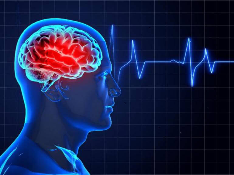
Symptoms of a Stroke
The stroke clinic consists of cerebral, meningeal (shell) and focal symptoms. The acute manifestation and rapid progression of the clinic is characteristic. Usually ischemic stroke has a slower development than hemorrhagic. Focal manifestations come to the fore from the onset of the disease, cerebral symptoms are usually mild or moderate, meningeal symptoms are often absent. Hemorrhagic stroke develops more rapidly, makes its debut with cerebral manifestations, against which focal symptoms appear and progressively increase. In the case of subarachnoid hemorrhage, meningeal syndrome is typical.
Cerebral symptoms are represented by headache, vomiting and nausea, impaired consciousness (stupor, stupor, coma). Approximately 1 out of 10 patients with hemorrhagic stroke have epipristup. The increase in cerebral edema or the volume of blood poured out during a hemorrhagic stroke leads to a sharp intracranial hypertension, mass effect and threatens the development of a dislocation syndrome with compression of the brain stem.
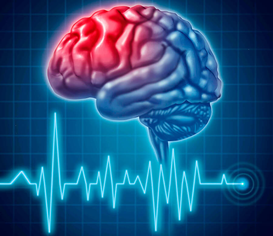
Focal manifestations depend on the location of the stroke. With a stroke in the pool of carotid arteries, central hemiparesis / hemiplegia occurs - a decrease / complete loss of muscle strength of the extremities of one side of the body, accompanied by an increase in muscle tone and the appearance of pathological stop signs. In the ipsilateral limbs, half of the face develops paresis of the facial muscles, which is manifested by a distortion of the face, lowering of the corner of the mouth, smoothing of the nasolabial folds, logophthalmos; when you try to smile or raise eyebrows, the affected side of the face lags behind the healthy or does not remain at all. These motor changes occur in the limbs and half of the face of the contralateral lesion of the side. In the same limbs, sensitivity decreases / falls out. Homonymous hemianopsia is possible - loss of the same half of the visual fields of both eyes. In some cases, photopsies and visual hallucinations are noted. Often there is aphasia, apraxia, decreased criticism, visual-spatial agnosia.
With a stroke in the vertebrobasilar basin, dizziness, vestibular ataxia, diplopia, visual field defects, dysarthria, cerebellar ataxia, hearing impairment, oculomotor disturbances, dysphagia are noted. Quite often, alternating syndromes appear - a combination of ipsilateral stroke of peripheral paresis of the cranial nerves and contralateral central hemiparesis. With a lacunar stroke, hemiparesis or hemigipesthesia can be observed in isolation.
Stroke diagnosis
Differential diagnosis of stroke
The primary task of diagnosis is the differentiation of stroke from other diseases that may have similar symptoms. The closed traumatic brain injury can be excluded by the absence of a traumatic history and external injuries. Myocardial infarction with loss of consciousness occurs as suddenly as a stroke, but there are no focal and cerebral symptoms, arterial hypotension is characteristic. A stroke manifesting a loss of consciousness and an epipristus can be mistaken for epilepsy. In favor of a stroke, the presence of a neurological deficit that accrues after paroxysm, the absence of epiproteins in the history, speaks.
At first glance, toxic encephalopathies are similar to stroke in acute intoxications (carbon monoxide poisoning, liver failure, hyper- and hypoglycemic coma, uremia). Their distinctive feature is the absence or weak manifestation of focal symptoms, often the presence of polyneuropathy, a change in the biochemical composition of blood corresponding to the nature of intoxication. Stroke-like manifestations can be characterized by hemorrhage in a brain tumor. Without the presence of an oncological history, it is clinically not possible to distinguish it from a hemorrhagic stroke. Intense headache, meningeal symptoms, nausea and vomiting with meningitis can resemble a picture of subarachnoid hemorrhage. In favor of the latter may indicate the absence of severe hyperthermia. A migraine paroxysm may have a picture similar to subarachnoid hemorrhage, however, it proceeds without sheath symptoms.
Differential diagnosis of ischemic and hemorrhagic stroke
After the diagnosis is established, the next step in the differential diagnosis is to determine the type of stroke, which is of paramount importance for the differential therapy. In the classic version, ischemic stroke is characterized by gradual progression without impaired consciousness in the opening, and hemorrhagic - by apoplektiform development with an early occurrence of a disorder of consciousness. However, in some cases, ischemic stroke can have an atypical onset. Therefore, in the course of diagnosis, one should rely on a set of various signs that testify in favor of a particular type of stroke.
So, for a hemorrhagic stroke, a history of hypertension with hypertensive crises is more typical, and for ischemic stroke, arrhythmia, valvular disease, myocardial infarction. The age of the patient also matters. The manifestation of the clinic during sleep or rest speaks in favor of ischemic stroke, and the beginning in the period of active activity in favor of hemorrhagic stroke. In most cases, the ischemic type of stroke occurs against the background of normal blood pressure, focal neurological deficit comes to the fore, arrhythmia, deafness of heart sounds is often noted. Hemorrhagic stroke, as a rule, makes its debut with elevated blood pressure with cerebral symptoms, shell syndrome and vegetative manifestations are often expressed, followed by the addition of stem symptoms.
Instrumental diagnosis of stroke
Clinical diagnosis allows a neurologist to determine the pool in which a vascular catastrophe occurred, to localize the focus of cerebral stroke, to determine its nature (ischemic / hemorrhagic). However, clinical differentiation of the type of stroke in 15-20% of cases is erroneous. A more accurate diagnosis is made possible by instrumental examinations. Optimal is an urgent MRI or CT scan of the brain. Tomography allows you to accurately determine the type of stroke, to clarify the location and size of the hematoma or ischemic focus, to assess the degree of cerebral edema and displacement of its structures, to detect subarachnoid hemorrhage or breakthrough of blood into the ventricles, to diagnose stenosis, occlusion and aneurysm of cerebral vessels.
Since there is not always the possibility of urgent neuroimaging, resort to lumbar puncture. Pre-conduct Echo-EG to determine / exclude the displacement of the middle structures. The presence of bias is a contraindication for lumbar puncture, which threatens in such cases the development of a dislocation syndrome. Puncture may be required when clinical data indicate subarachnoid hemorrhage, and tomographic methods do not detect blood accumulation in the subarachnoid space. In ischemic stroke, cerebrospinal fluid pressure is normal or slightly increased, the study of cerebrospinal fluid does not reveal significant changes, a slight increase in protein and lymphocytosis can be determined, in some cases a small admixture of blood. With hemorrhagic stroke, there is an increase in cerebrospinal fluid pressure, a bloody color of cerebrospinal fluid, a significant increase in protein concentration; in the initial period, unchanged red blood cells are determined, later - xantochromic.
Doppler ultrasonography of extracranial vessels and transcranial Doppler ultrasound diagnose angiospasm and occlusion, determine the degree of stenosis and evaluate collateral circulation. Emergency angiography of the brain is necessary to address the issue of the appropriateness of thrombolytic therapy, as well as for the diagnosis of aneurysms. Preference is given to MRI angiography or CT of cerebral vessels. In order to identify the causes of stroke, an ECG, echocardiography, a clinical blood test with platelet count, a coagulogram, a biochemical blood test (including blood sugar), a urinalysis, and a blood gas composition are performed.
Stroke treatment
The optimal duration of hospitalization and initiation of therapy are the first 3 hours from the debut of clinical manifestations. Treatment in the most acute period is carried out in intensive care units of specialized neurological departments, then the patient is transferred to the early rehabilitation unit. Before the type of stroke is established, basic undifferentiated therapy is carried out, after an accurate diagnosis is made, specialized treatment is followed, and then a long rehabilitation.
Undifferentiated treatment of stroke includes correction of respiratory function with pulse oximetric monitoring, normalization of blood pressure and cardiac activity with daily monitoring of ECG and blood pressure (together with a cardiologist), regulation of homeostatic parameters (electrolytes and blood pH, glucose level), the fight against cerebral edema (osmodiuretics, corticosteroids hyperventilation, barbiturate coma, cerebral hypothermia, decompressive trepanation of the skull, external ventricular drainage).
In parallel, symptomatic therapy is carried out, which may consist of hypothermic agents (paracetamol, naproxen, diclofenac), anticonvulsants (diazepam, lorazepam, valproates, sodium thiopental, hexenal), antiemetics (metoclopramide, perphenazine). With psychomotor agitation, magnesium sulfate, haloperidol, barbiturates are indicated. Basic stroke therapy also includes neuroprotective therapy (thiotriazolin, piracetam, choline alfoscerate, glycine) and the prevention of complications: aspiration pneumonia, respiratory distress syndrome, pressure sores, uroinfection (cystitis, pyelonephritis), pulmonary embolism, thrombophlebitis.
Differentiated treatment of stroke corresponds to its pathogenetic mechanisms. In ischemic stroke, the main is the speedy restoration of blood flow of the ischemic zone. For this purpose, drug and intra-arterial thrombolysis using tissue plasminogen activator (rt-PA), mechanical thrombolytic therapy (ultrasound thrombus destruction, thrombus aspiration under tomographic control) are used. With the proven cardioembolic genesis of stroke, anticoagulant therapy with heparin or nadroparin is performed. If thrombolysis is not indicated or cannot be carried out, then antiplatelet drugs (acetylsalicylic acid) are prescribed. In parallel, vasoactive drugs (vinpocetine, nicergoline) are used.
The priority in the treatment of hemorrhagic stroke is to stop bleeding. Hemostatic treatment can be carried out with calcium preparations, vicasol, aminocaproic acid, ethamzilate, aprotinin. Together with a neurosurgeon, a decision is made on the appropriateness of surgical treatment. The choice of surgical tactics depends on the location and size of the hematoma, as well as on the condition of the patient. Possible stereotactic aspiration of the hematoma or its open removal by trepanation of the skull.
Rehabilitation is carried out using regular courses of nootropic therapy (nicergoline, pyritinol, piracetam, ginkgo biloba, etc.), physical therapy and mechanotherapy, reflexology, electromyostimulation, massage, physiotherapy. Patients often have to re-establish motor skills and learn self-care. If necessary, psychiatric specialists and psychologists conduct psychocorrection. Correction of speech disorders is carried out by a speech therapist.
Stroke Prediction and Prevention
The lethal outcome in the 1st month with ischemic stroke varies from 15 to 25%, with hemorrhagic stroke - from 40 to 60%. Its main causes are edema and dislocation of the brain, the development of complications (pulmonary embolism, acute heart failure, pneumonia). The greatest regression of neurological deficit occurs in the first 3 months. a stroke. Often there is a worse recovery of movements in the arm than in the leg. The degree of restoration of lost functions depends on the type and severity of the stroke, the timeliness and adequacy of the provision of medical care, age, and related diseases. After a year from the moment of a stroke, the likelihood of further recovery is minimal, after such a long period usually only aphasia is amenable to regression.
The primary prevention of stroke is a healthy diet with a minimum amount of animal fats and salt, a mobile lifestyle, balanced and calm nature, avoiding acute stressful situations, and the absence of bad habits. The prevention of both primary and recurrent stroke is facilitated by the effective treatment of cardiovascular pathology (blood pressure correction, treatment of coronary heart disease, etc.), dyslipidemia (taking statins), and reduction of overweight. In some cases, the prevention of stroke is surgery - carotid endarterectomy, reconstruction of the vertebral artery, the formation of extra-intracranial anastomosis, surgical treatment of AVM.